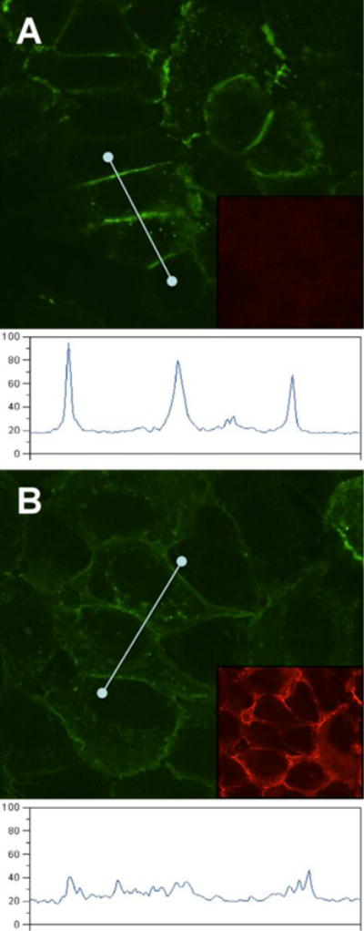
Figure 3.

Redistribution of GFP-nectin-1 from junction of epithelial cells. A. detection of GFP-nectin-1a accumulating at junctions between human ECC-1-NIG cells. B. After incubation with soluble gD(285t) (1 μM) for 2h, the intensity of GFP at cell junctions is decreased. Under each condition, an intensity profile following a straight line through several cell junctions is shown. Images were captured under similar conditions. Intensity units are arbitrary. The insets show gD immunostaining with polyclonal serum R7 followed by Alexa594-coupled secondary antibody.
