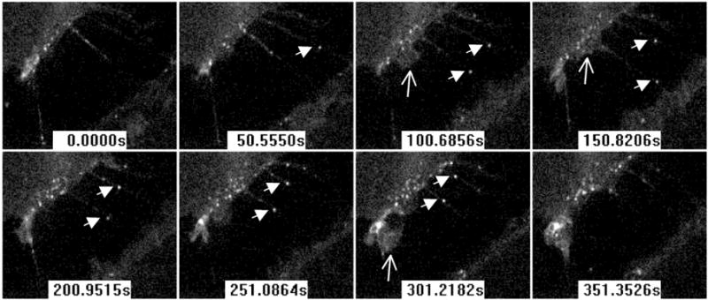
Figure 7.

HSV surfing on filopodia of cells expressing GFP-nectin-1 without cytoplasmic tail. HSV-K26 with GFP-tagged capsid is added to B78-NGC-389 cells immediately prior to recording. Confocal images show the space between two cells connected by filopodia. Filled arrows indicate two moving capsids on two different filopodia. Open arrows indicate areas of ruffling membrane. Time from the start of recording is indicated in seconds.
