
Fig. 1.
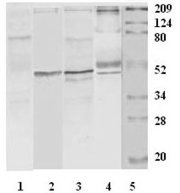
The Western blot patterns of the immunoreactive proteins in Vero-E6 cells infected by the SARS-CoV. Each lane was loaded with 50 mg of the protein from the lysate of Vero-E6 cells. Lane 1: control, the serum from a healthy donor as the primary antibody; Lane 2–4: SARS-CoV, the sera from SARS patients as primary antibodies; Lane 5: standard protein markers.