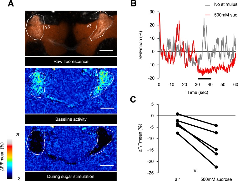
Fig 3. Sugar reward suppresses the baseline activity of PAM-γ3.
(A) Representative raw fluorescence in the PAM-γ3 neurons of the MB441B-GAL4/UAS-GCaMP5 fly with the mushroom bodies labelled with dsRed (orange). Two-photon images for one second before (baseline) and during the first second of sucrose stimulation (during sugar) are averaged and presented in a pseudocolor code. Region of interest is defined by terminals of PAM-γ3 in the medial lobes (outlined). Scale bar, 20 μm. (B) Time course of baseline activity (gray) and sugar response (red). Black bar: stimulus application. (C) Average calcium responses of PAM-γ3 neurons. Sucrose ingestion significantly reduces the activity level of PAM-γ3 (Wilcoxon matched-pairs signed rank test, n = 5). * p < 0.05.

