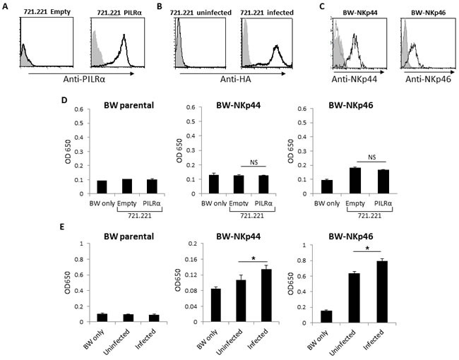
Figure 2. PILRα expressing cells does not increase IL2 secretion of BW-NKp44 and BW-NKp46 cells.

A. FACS staining of 721.221 cells transfected either with an empty vector as control, or with PILRα. Grey filled histograms are background control staining. Black line histograms represent specific anti-PILRα staining. B. FACS staining of 721.221 cells in the presence or absence of PR8 Influenza. Grey filled histograms are background control staining. Black line histograms represent specific anti-HA staining. C. FACS staining of parental or NKp44 and NKp46 transfected BW cells. Grey filled histograms are background control staining. Grey line histograms represent the staining of the parental BW cells with the appropriate mAbs, black line histograms represent staining of NKp44/46 transfected BW cells with the appropriate mAbs. D. IL2 secretion from parental BW (left), BW-NKp44 (middle) and BW-NKp46 (right) cells. IL2 secretion was measured by ELISA (OD 650nm) following incubation with 721.221 cells transfected either with an empty vector or with PILRα. E. IL2 secretion of parental BW (left), BW-NKp44 (middle) and BW-NKp46 (right) cells. IL2 secretion was measured by OD 650nm following incubation with 721.221 in the presence (designated infected) or in the absence (designated uninfected) of influenza PR8. Figure show combine 3 independent experiments. *p < 0.05, NS-not significant. Statistics was performed using student T-test.
