
Figure 2. M1.HpyAVI exists as dimer in crystal and solution.
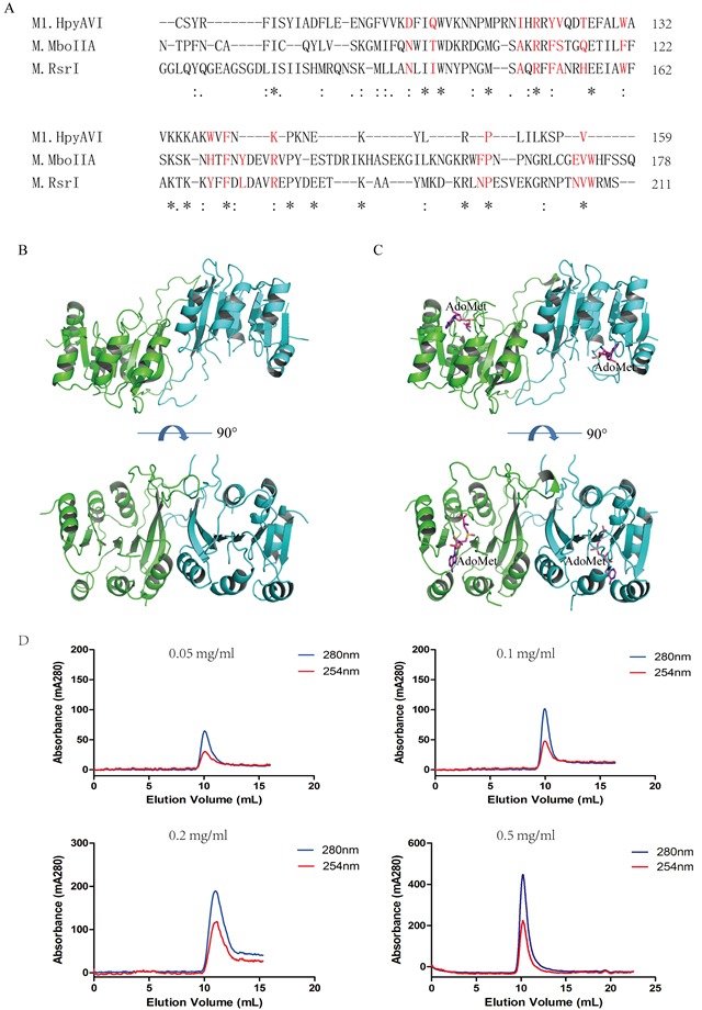
A. A conserved interface area of β-class MTases is defined in M1.HpyAVI. Residues that involved are signed in red color; Dimerization of free-form M1.HpyAVI B. and cofactor-bound M1.HpyAVI C. The two monomers are marked in green and blue, AdoMet molecules are marked in magenta. D. Gel-filtration analysis revealed that M1.HpyAVI exist as a dimer in solution. FPLC system coupled to a Superdex 75 10/300 column. Elution profiles at 280 nm (blue) and 260 nm (red) are: different concentration (0.05, 0.1, 0.2, 0.5 mg/ml) of M1.HpyAVI protein.