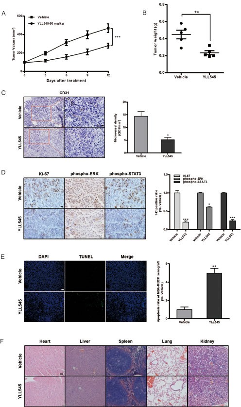
Figure 6. YLL545 exerts antitumor activities in vivo.

A. and B. A total of 2.5×10⁷ MDA-MB-231 cells were injected into the fat pad of nude mice (n = 5). Tumor development was monitored for 14 days. When tumors reached a volume of about 100 mm3, the mice were treated with 50 mg/kg/d YLL545 or vehicle for another 12 days. Cross-sectional diameters of tumors from YLL545- and vehicle-treated mice were measured. Approximate tumor volumes (A) and weights (B) were calculated as described in Materials and Methods. **P < 0.01 and ***P < 0.001 vs respective control in Student's t-test. C. Expression of CD31 in breast cancer xenografts was examined by immunohistochemical staining. MVD was determined by CD31 staining. MVD was counted in five random fields in each tumor. *P < 0.05 vs respective control in Student's t-test. Scale bars, 20 μm. D. Ki67, phospho-STAT3 and phospho-ERK1/2 levels in breast cancer xenografts were examined by immunohistochemical staining. *P < 0.05 and ***P < 0.001 vs respective control in Student's t-test. Scale bars, 20 μm. E. YLL545-induced tumor cell apoptosis was measured by TUNEL staining in YLL545- and vehicle-treated mice. **P < 0.01 vs respective control in Student's t-test. Scale bars, 100 μm. F. YLL545 did not cause obvious pathologic abnormalities in normal tissues. H&E staining of paraffin embedded sections was performed on heart, liver, spleen, lung, and kidney tissues from YLL545- and vehicle-treated mice. Scale bars, 50 μm.
