
Figure 4. Andrographolide induced apoptosis signaling is dependent on ER Stress.
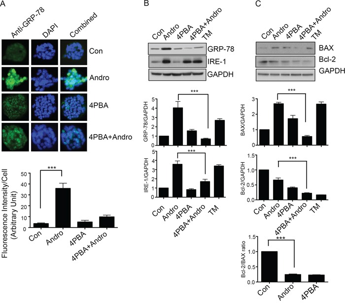
A. T84 cells were grown on coverslips and treated with Andrographolide IC50 in the presence or absence of 4-PBA and GRP-78 expression was evaluated by immunofluorescent staining. Nuclei were stained using DAPI and cells were examined by fluorescence microscopy. Fluorescence intensity was determined and compared with untreated T84 cells. B. T84 cells treated with Andrographolide IC50 for 48 h were lysed and protein expression was determined by immunoblotting for GRP-78, IRE1, and GAPDH. Densitometry analysis was performed and normalized with GAPDH expression to demonstrate significant reductions in expression of GRP-78, IRE1, in the present of 4-PBA. C. Cell lysates were analyzed for pro-apoptotic BAX and anti-apoptotic Bcl-2 expression by immunoblot analysis and quantified by densitometry. The lower graph shows the ratio of Bcl-2/BAX. The results shown are from three independent experiments. (***P < 0.001)