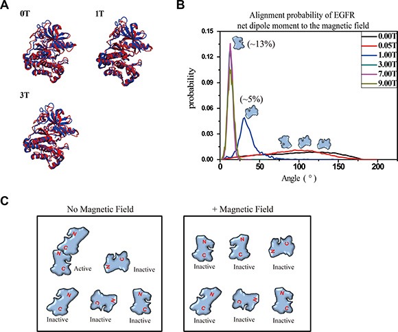
Figure 5. Molecular dynamics simulation shows that 1-9T SMFs affect orientation of EGFR-KD.

(A) The superimposed models between crystal structures (blue) of EGFR-KD and the final snapshot of MD under different SMFs (red). (B) Probability distribution of the angles between the net dipole moment of EGFR-KD and field direction. (C) Cartoons illustrate the effect of SMF on protein alignment and activation.
