
Figure 6. Murine monocytes and macrophages express the functional TRAIL receptor DR5.
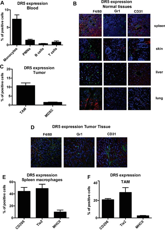
A. Flow cytometry analysis of murine TRAIL-R (DR5) in blood monocytes, granulocytes, T and B lymphocytes; results are shown as % of positive cells relative to total CD45+ cells (mean±SE of 29 blood samples). B. Immunofluorescence analysis of DR5, macrophages (F4/80), granulocytes (Ly6G) and endothelial cells (CD31) in normal murine tissues, (DR5 in red, F4-80/Ly6G/CD31 in green and nuclei in blu). C. DR5 expression in TAM and in MDSC from tumor tissues; results are shown as % of positive cells (Mean ± SD of 3 experiments); D. immunofluorescence analysis of DR5 in fibrosarcoma tumor tissues (DR5 in red, F4-80/Ly6G/CD31 in green and nuclei in blu). E-F. DR5 expression in CD206+, Tie2+, MHCII+ macrophages from spleen (E) and tumor (F); results are shown as % of positive cells (Mean ± SD of 3 experiments). Statistical analysis: *P < 0.05, ** P < 0.01, *** P < 0.001 (Student's t test).