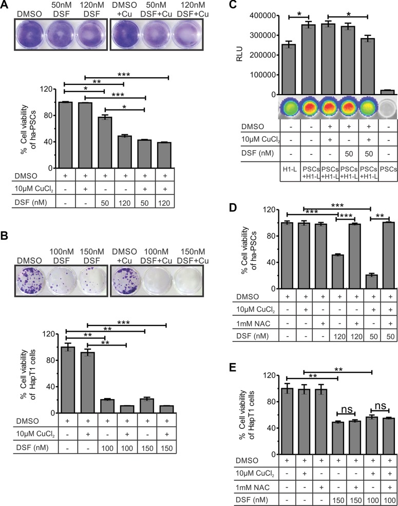
Figure 5. Effect of DSF and/or CuCl2 on the viability of hamster-PSCs and HapT1 cancer cells.
(A) After 24 hours of treatment with DSF or DSF+Cu or vehicle control, the ha-PSCs were cultured with normal culture medium. On fifth day of culture, the plates were processed for crystal violate staining and quantification. The data clearly show the significant cytotoxic effect of 120 nM DSF (with and without 10 μM CuCl2) and 50 nM DSF + 10 μM CuCl2 on ha-PSCs. The figure shows representative images of the crystal violate stained plates (one out of three independent experiments in duplicates) and its corresponding quantified values (n = 3). (B) The colony forming assay using HapT1 cancer cells shows the cytotoxic effect of DSF alone and in combination with Cu (24 hours of treatment followed by 5 days of culture). Representative image of a crystal violet-stained plate (one out of three independent experiments in duplicates) is shown. Data obtained from the crystal violate quantification corresponds to the number of viable cells (n = 3). (C) Co-culture of ha-PSCs and HapT1-Luc cancer cells (H1-L) shows the role of PSCs in the growth of cancer cells. However, pre-treatment of PSCs with DSF+Cu significantly reduced the growth advantage conferred by PSCs to H1-L cells. D & E) Cell viability checked through MTT assay showed the rescue of ha-PSCs form DSF-mediated cell death after pre-incubation with 1 mM NAC; however, HapT1 cells did not get benefit from NAC pre-incubation. Data presented as mean ± SE; *p < 0.05; **p < 0.005; ***p < 0.0005.

