
Figure 1. A. Individual perivascular cells express different markers.
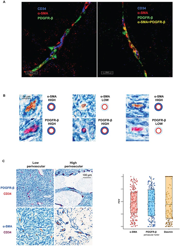
The triple staining with both perivascular cell markers (PDGFR-β (red) or α-SMA (green)) and CD34 (blue) demonstrate presence of perivascular cells of three distinct perivascular cell types: PDGFR-β- and α-SMA-single positive cells and double-positive cells. B. Individual vessels are characterized by relatively independent perivascular expression of PDGFR-β and α-SMA. Serial sections of three vessels stained with α-SMA (blue staining, upper panels) or PDGFR-β (blue staining, lower panel) and CD34 (brown or red staining) showing independent perivascular expression of PDGFR-β and α-SMA. Note in left part an α-SMA-high / PDGFR-β-high vessel, in the middle part an α-SMA-low / PDGFR-β –high vessel and in right an α-SMA-high / PDGFR-β-low vessel. C. Inter-tumoral variation of perivascular marker expression. Left panel: Representative images of PDGFR-β and α-SMA (blue) double staining with CD34 (red or brown) showing areas of low or high perivascular marker expression. Note different intensity of perivascular staining (blue) in different cases. Right panel: Distribution of the fraction of covered vessels with PDGFR-β, α-SMA and Desmin in Nordic-VII cohort (right panel). The cases of the cohort are characterized by heterogeneous perivascular cell coverage defined by three markers.