
Figure 5. Blockade of IL-8 signaling reverts erlotinib resistance.
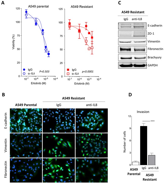
A. Assessment of erlotinib response in parental vs. erlotinib-resistant A549 cells previously treated with control IgG or neutralizing anti-IL-8 antibody [p values calculated by two-way ANOVA]. B. Immunofluorescent analysis (green signal) of epithelial E-cadherin and mesenchymal vimentin and fibronectin in A549 parental vs. erlotinib-resistant cells treated with a control IgG or a neutralizing anti-IL-8 antibody. Blue signal corresponds to DAPI stained nuclei. C. Western blot analysis of indicated markers in A549 erlotinib-resistant cells treated with control IgG or anti-IL-8 antibody. D. Invasion assay performed with indicated cells treated with control IgG or anti-IL-8 antibody prior to the assay.