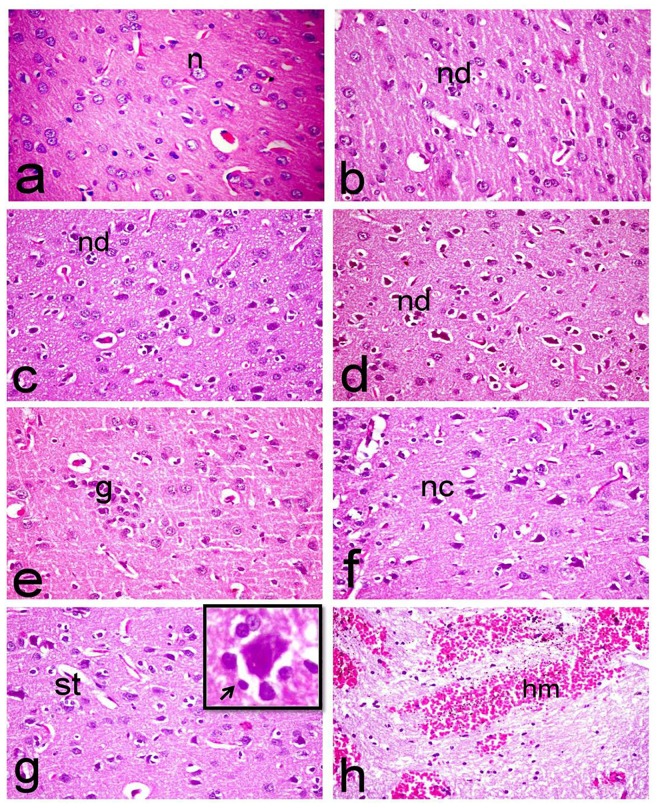
Fig 5. Histopathological investigation of brain tissues.
Brain sections of (a) normal rats showing normal morphology of cerebral cortex with normal rounded neuronal cells (n), (b) ipPDC-treated rats showing gliosis and degeneration of individual neuronal cells associated with neuronophagia, (c) inPDC (0.5 mg/kg)-treated rats showing wide spread neuronal cell degeneration (nd) with neuronophagia of the degenerated neurons as well as vacuolation of the neuropil, (d & e) inPDC (1 mg/kg)-treated rats showing (d) neuronal cell degeneration (nd) associated with neuronophagia and neuronal loss, (e) focal gliosis (g), and (f, g & h) inPDC (2 mg/kg)-treated rats showing (f) neuronal cell necrosis (nc) with intensely eosinophilic shrunken neuronal cell bodies, (g) proliferation of glia cells, satellatosis (st), and neuronophagia (insert) in which the degenerated neuron is surrounded by astrocytes and microglia cells (arrow) (h) extensive hemorrhage (hm) in the white matter. (H&E, X40).

