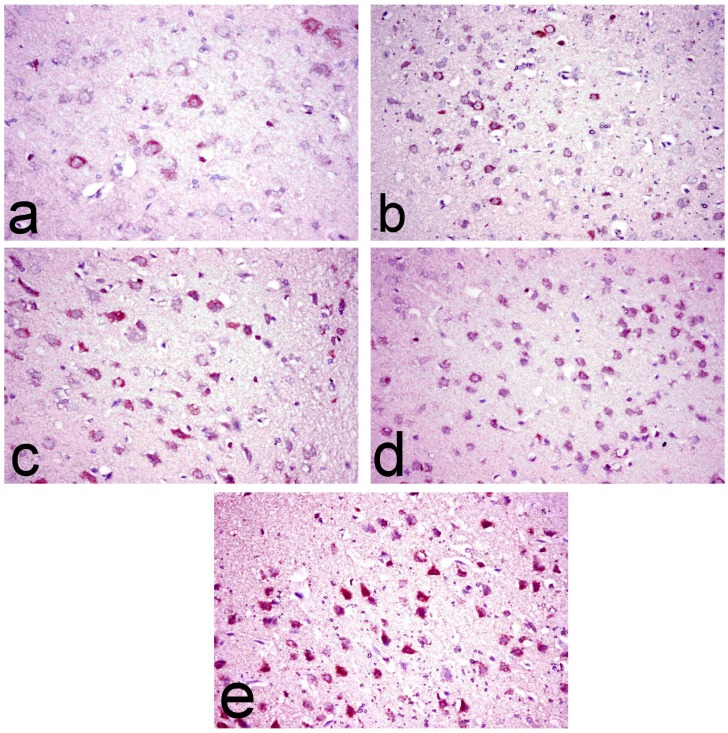
Fig 7. Immunohistochemical investigation of brain tissues.
Brain sections of (a) normal rats showing scattered individual COX-2 immune-stained cells, (b) ipPDC-treated rats showing an increase of COX-2 immune-stained cells with perinuclear immunereactivity, (c) inPDC (0.5 mg/kg)-treated rats showing COX-2 immune-stained cells with perinuclear immunereactivity, (d) inPDC (1 mg/kg)-treated rats showing significant increase of COX-2 immune-stained cells, and (e) inPDC (2 mg/kg)-treated rats showing numerous intensely brown COX-2 immune-stained cells distributed all over the cerebral cortical layer. (COX-2 immunohistochemical staining, X40).

