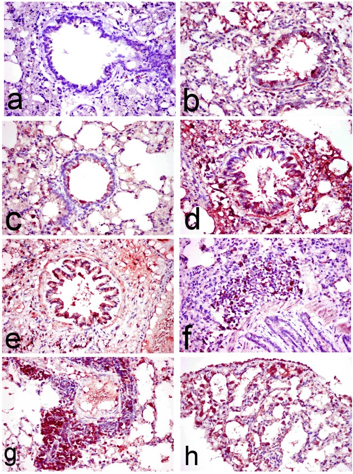
Fig 8. Immunohistochemical investigation of lung tissues.
Lung sections of (a) normal rats showing no COX-2 immune-stained cells, (b) ipPDC-treated rats showing COX-2 immune stained cells lining the bronchiolar mucosa and alveolar wall, (c) inPDC (0.5 mg/kg)-treated rats showing COX-2 immune-stained cells lining the bronchiolar mucosa and alveolar wall as well as in the alveolar lumina, (d) inPDC (1 mg/kg)-treated rats showing abundant COX-2 immune-stained cells lining the bronchiolar wall and the peribronchiolar tissue, and (e, f, g & h) inPDC (2 mg/kg)-treated rats showing (e) COX-2 immune-stained cells that appeared intensely brown lining the bronchiolar wall, (f) COX-2 immune-stained cells in the peribronchial tissue, (g) COX-2 immune-stained cells in the perivascular tissue, and (h) COX-2 immune-stained cells in the alveolar lumen. (COX-2 immunohistochemical staining, X40).

