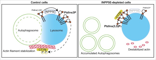
Figure 1.
Model of INPP5E-mediated autophagosome-lysosome fusion in neuronal cells. In control cells (left), INPP5E is transiently localized on lysosomes, where it dephosphorylates PtdIns(3,5)P2. A decrease in PtdIns(3,5)P2 level and an increase in PtdIns3P level on lysosomes lead to activation of CTTN/cortactin, which can then bind and stabilize actin filaments. The stabilized actin filaments, in turn, facilitate autophagosome-lysosome fusion. In INPP5E-depleted cells (right), CTTN is trapped by elevated levels of PtdIns(3,5)P2, which accumulates on lysosomes in the absence of INPP5E function, and remains inactive, leading to destabilization of actin filaments. Enclosed autophagosomes thus accumulate in INPP5E-depleted cells.

