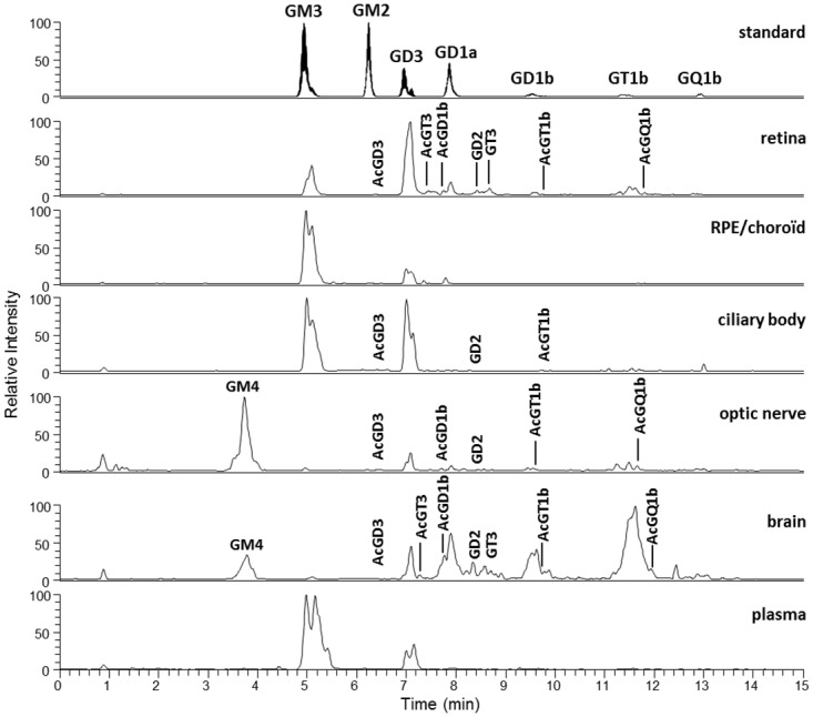
Fig 3. Precursor ion chromatograms of the standard mixture, retina and other ocular tissues, brain and plasma.

A representative sample of each tissue was obtained by pooling an aliquot of every GG extract samples (4–7) for each tissue type. The QqQ mass spectrometer was operated in the precursor ion scanning of the characteristic fragment of GG, N-Acetylneuraminic acid, at m/z 290, in the negative ionization mode.
