
Figure 1. Ectosomes have a unique protein composition.
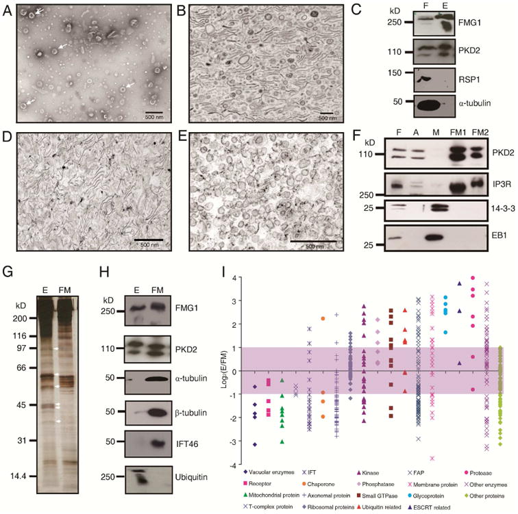
A) Negative stained isolated ectosomes (arrows) released from flagella of vegetative cells. B). Electron micrograph of thin sectioned, isolated ectosomes. C). Immunoblot of flagella (F) and ectosomes (E) with antibodies against flagellar membrane proteins (FMG1 and PKD2) and axonemal proteins (RSP1 and α-tubulin). D). Electron micrograph of flagellar membrane (FM1) isolated with 1% NP-40. E). Electron micrograph of flagellar membrane vesicles (FM2) recovered from the supernatant after removing the detergent. F).Western blot of 10 ug of protein from flagella (F), axoneme (A), matrix (M), FM1 and FM2 with antibodies against PKD2, IP3R (IP3 receptor), 14-3-3 and EB1. Membrane proteins are enriched in FM1 and FM2. G). Silver stained SDS-PAGE gel of proteins from ectosomes (E) versus flagellar membrane (FM). Arrows indicate the proteins enriched in ectosomes. H). Western blots of the same amount of protein of ectosomes (E) and flagellar membrane (FM) with antibodies recognizing the indicated proteins. I). Comparison of the proteins in ectosomes versus in the isolated flagellar membrane (FM1+FM2) using iTRAQ analysis. In total, 563 proteins with more than 3 peptides were categorized into 19 subgroups according to their known or putative function. Proteins with the Log2(E/FM) > 1 were considered as ectosome enriched, see in Table S1; -1 < Log2(E/FM) < 1 were considered to be equal in ectosomes and flagellar membranes; and Log2(E/FM) < -1 were considered to be enriched in the flagellar membrane, see in Table S2. Membrane proteins present in ectosome and flagellar membrane are shown in Table S3. Comparison of flagellar membranes isolated with detergent and freeze/thaw are shown in Figure S1.