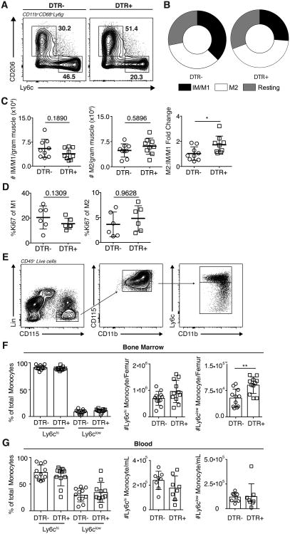
Figure 6. Treg depletion is accompanied by shift toward increased M2.
Systemic depletion of Tregs following DT treatment of DTR transgenic mice. (A) Representative FACS plots, (B) distribution and (C) absolute number and fold change of IM/M1 and M2 one day following final DT treatment. (D) Effect of Treg depletion on skeletal muscle M1 and M2 proliferation (Ki67+) one day following final DT treatment. (A-D) Results are representative of n = 10 per group from four experiments. (E) Gating scheme for Ly6chi and Ly6clow monocytes. Frequencies and absolute numbers of (F) bone marrow and (G) blood Ly6chi and Ly6clow monocytes (CD45+TCRβ-Ly6G-SiglecF-NK1.1-TER119-CD11b+CD115+) one day following final DT treatment. Results are representative of n = 13 per group from three experiments; error bars are the SD. *P < 0.05; **P < 0.01; (C, E, F) Kruskal-Wallis test, (D-F) Student's t test.

