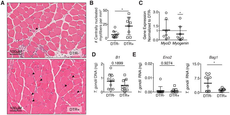
Figure 7. Treg ablation rescues skeletal muscle fiber regeneration.
(A) Representative H&E stains of hindlimb muscles one day following final DT treatment in DTR- and DTR+ mice 28 dpi. Arrows indicate centrally nucleated myofibers. (B) Quantification of regenerating fibers (centrally nucleated myofibers) in muscle sections per mm2 (C) Fold expression of myogenic targets MyoD and myogenin in infected DTR+ skeletal muscle normalized to DTR- skeletal muscle following DT treatment Statistics were calculated on log-transformed fold change values. (D) qRT-PCR quantification of total parasite burden by B1 and (E) parasite stage specific transcripts Bag1 bradyzoite (left) and Eno2 tachyzoite (right) following DT treatment. Results are representative of n = 10 per group from four experiments; error bars are the SD. *P < 0.05; Student's t test

