Figure 1.
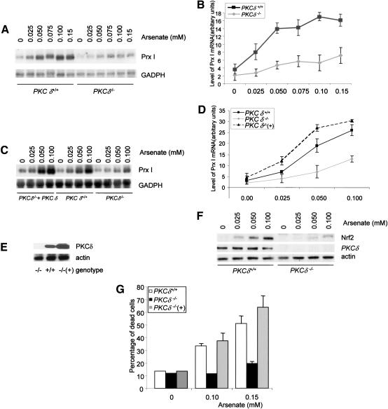
Induction of Prx I in PKC δ deficient cells is reduced. (A) PKC δ-deficient and wild-type control MEFs were treated with different concentrations of sodium arsenate for 6 h. Total RNA was isolated from these cells and was analyzed by Northern blot using a radiolabeled Prx I probe. (B) Quantitation data from three experiments. (C) PKC δ-deficient osteoblasts showed compromised induction of Prx I, which was rescued by reconstitution of PKC δ. The experiments were done as described in A. (D) Quantitation data from three experiments. (E) Western blot shows that PKC δ was expressed by a retroviral vector in PKC δ-deficient osteoblasts. (F) Up-regulation of Nrf2 is suppressed in PKC δ-deficient cells. Mutant and wild-type control osteoblasts were treated with different concentrations of arsenate for 6 h and collected. Expression of Nrf2 was analyzed by Western blot using anti-Nrf2 antibodies. Actin is used as a loading control. Expression of PKC δ was detected using a monoclonal antibody. (G) PKC δ-deficient osteoblasts were resistant to the cytotoxic effect of arsenate. Mutant and wild-type osteoblasts were treated with different concentrations of arsenate for 20 h and cell death rates were determined by the trypan blue exclusion method.

