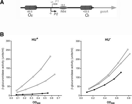
Figure 1.
(A) Schematic structure of the P2∼gusA fusion used to study looping repression. In this construct the gal regulatory region contains the external (OE) and internal (OI) operator sites, the P2 promoter, and the HU-binding site hbs. The P1 promoter is inactivated by a point mutation. The transcription start site +1 of P1 is used as a reference in the numbering system. (B) Repression of the P2 promoter in vivo. Differential rates of β-glucuronidase synthesis from P2∼gusA in hup+ DM0022 (left panel) and in ΔhupAΔhupB strain DM0100 (right panel) cells in the presence of plasmid carrying galR+ (filled squares), galRH327R (open circles), and galRT322R (open squares).

