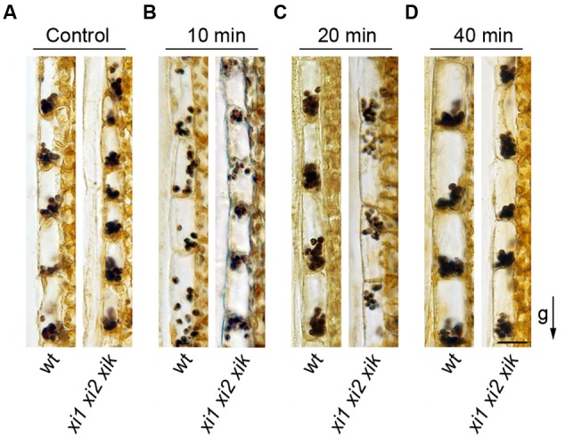
FIGURE 8.

Amyloplast distribution in endodermal cells. (A) Localization of amyloplasts in the stems of control plants. (B–D) Inflorescence stems were reoriented 180° and gravistimulated for (B) 10, (C) 20, or (D) 40 min. The arrow indicates direction of gravity (g). Scale bar 20 μm.
