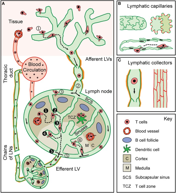
Figure 1.
T cell traffic through the lymphatic vascular system. (A) Recirculating effector-memory T cells in peripheral tissues ➀ enter afferent lymphatic vessels (LVs). The exact point of entry or the mode of intralymphatic movement has not been investigated so far. T cells that ➁ arrive in the lymph node (LN) subcapsular sinus (SCS) have been shown to cross the lymphatic endothelium into the LN parenchyma at the level of the ➂ SCS or of the ➃ medullary sinuses. Some T cells do not enter the LN parenchyma but ➄ directly exit through the efferent LV located at the hilus region of the LN. Recirculating naïve and central memory T cells arrive in the LN either via the blood (high endothelial venules) or via the afferent LV draining from an upstream LN (i.e., efferent lymph). ❶ T cells within the LN ❷ make random contact with the sinuses before entering and ❸ actively crawling or passively flowing within the sinuses. T cells were observed to ❹ cross the sinuses several times before finally being ❺ passively carried away into the efferent LV. T cells in the efferent LV circulate through downstream LNs before being returned to the blood circulation via the thoracic duct. (B) Lymphatic capillaries are composed of oak leaf-shaped lymphatic endothelial cells (LECs), which partially overlap and are held together by button-like associated junctional adhesion molecules (red lines). This setup creates open flaps through which leukocytes, fluid, and macromolecules enter into the vessel lumen. (C) LECs in collecting vessels have a cuboidal shape and are connected by continuous cell-cell junctions (red lines). Collecting vessels contain intraluminal valves and are surrounded by a basement membrane and contracting smooth muscles cells (orange).

