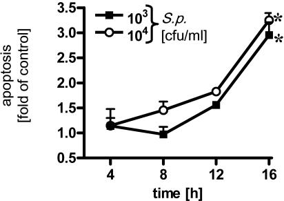
FIG. 7.
Time- and dose-dependent apoptosis of pneumococcus-infected human bronchial epithelial cells. BEAS-2B cells were infected for different times with S. pneumoniae (S.p.) (103 and 104 CFU/ml), and DNA fragmentation was measured. An asterisk indicates that the P value was <0.05 for a comparison with the unstimulated control at a single time.

