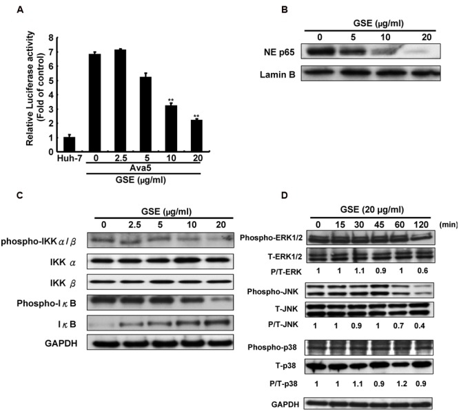
FIGURE 3.

Grape seed extract reduced NF-κB transactivity and MAPK phosphorylation for suppression of COX-2 expression in HCV replicon cells. (A) GSE reduced NF-κB transactivity in Ava5 cells. Ava5 cells were transiently transfected with pNF-κB-Luc, which contained an NF-κB binding element linked firefly luciferase reporter gene. The pNF-κB-Luc-transfected cells were treated with 20 μg/ml of GSE for 3 days. Subsequently, the extracted lysates of transfected cells were analyzed by luciferase activity assay. The relative NF-κB transactivity was presented as fold changes compared to parental Huh-7 cells in which luciferase activity was presented as 1. The GSE treatment downregulated (B) NF-κB phosphorylation and (C) the HCV-induced NF-κB signaling pathway. Ava5 cells were treated with GSE in different concentrations (0–20 μg/ml) for 3 days and the nuclear lysates were isolated as described in Section “Materials and Methods.” The nuclear translocation of NF-κB were analyzed by Western blotting with anti-phospho-p65 and anti-Lamin B (loading control) antibodies. The effects of GSE on NF-κB regulation were analyzed by Western blotting with various antibodies against IKKα, phospho-IKKα/β, IκB-α, phospho-IκB-α, and GAPDH (loading control). (D) GSE treatment reduced the phosphorylation level of ERK and JNK. Ava5 cells were treated with 20 μg/ml of GSE and the lysates extracted at the indicated time points after the treatment. The protein expressions were analyzed by Western blotting with antibodies against MAPK (ERK1/2, p38, and JNK), phospho-MAPK (p-ERK1/2, p-p38, and p-JNK), and GAPDH (loading control). Data are represented as the mean ± SD for three independent experiments. ∗P < 0.05; ∗∗P < 0.01.
