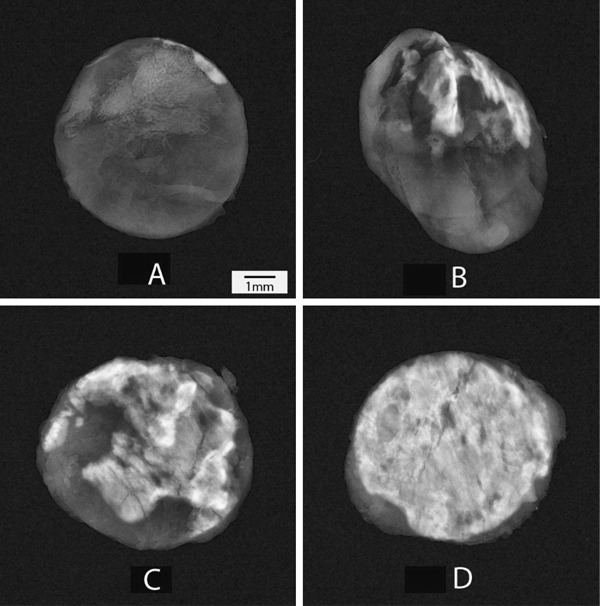
Fig. 2.

Representative X-ray images of tissue discs after explant of test group (a), porcine tissue treated with AOA (b), bovine pericardial tissue treated with Linx (c) and bovine pericardial tissue treated with glutaraldehyde only (d). The white area represents the calcified nodule. The X-ray images were sorted based on the calcium content and the median calcium images were selected as representative
