Figure 3.
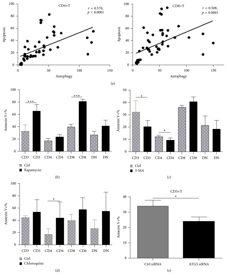
T cells with increased autophagy following stimulation were prone to apoptosis. Autophagy and apoptosis of T cells from SLE patients were detected after stimulation with anti-CD3/C28 antibodies for different time. And their correlation was analyzed. Apoptosis was also measured after autophagy activation with rapamycin (50 ng/mL, 72 h, simultaneously with anti-CD3/28 stimulation), or suppression with 3-methyladenine (3-MA) (5 mM, 6 h, following anti-CD3/28 stimulation for 48 h) or chloroquine (CQ, 50 μM for 24 h, following anti-CD3/28 stimulation for 48 h). In vitro experiments were performed in triplicate. (a) Correlation of apoptosis of both CD4+T and CD8+T cells from SLE patients with autophagy level following anti-CD3/28 stimulation (n = 32). (b) Activation of autophagy with rapamycin further promote apoptosis of T cells, especially in CD8+T cells (n = 7). (c) Suppression of autophagy with 3-MA rescued T cells (mainly CD4+T) from apoptosis (n = 7). (d) Treatment with chloroquine increased apoptosis of CD4+T cells (n = 6). (e) ATG5 silencing decrease apoptosis of T cells significantly. ∗ p < 0.05; ∗∗∗ p < 0.001.

