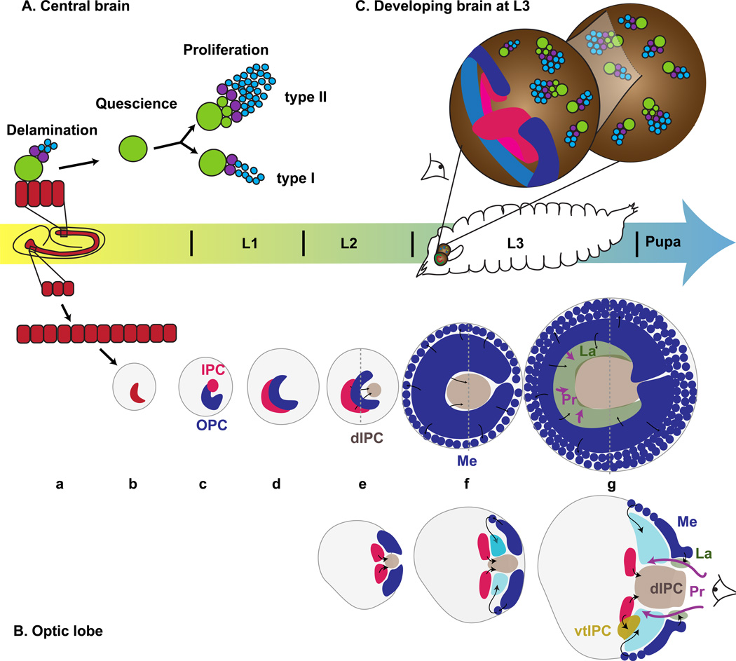
Figure 2. The development of the optic lobe of Drosophila.
(A) Central brain: Neuroblasts (green) delaminate from the neuroepithelium (red) during the embryonic stages and generate GMCs (purple) and larval neurons (blue) before they become quiescent. At L1, Type I and Type II neuroblasts are reactivated and exhibit different modes of proliferation to generate adult neurons. (B) Optic lobe: a) Cells in the procephalic regions generate the optic placode neuroepithelium (red), which invaginates (b); c) The optic placode split into inner proliferation center (IPC, pink) and outer proliferation center (OPC, blue) and adopt a crescent shape (d). e) Cells migrate out of the IPC to form the dIPC (light brown). f) The medial edge of the OPC becomes neuroblasts that will produce the medulla (Round blue cells). g) The inner OPC generates Lamina progenitors (La, green) that will produce lamina neurons upon induction by photoreceptors (Pr, pink arrows). The ventral tip of the IPC generates lobula complex neurons (vIPC, dark orange). Lower diagrams are cross sections at the dashed line in upper diagrams. (C) 3D representation of the brain at L3. Central brain neuroblasts and their progeny, OPC (blue) and IPC (pink). The eye symbol shows the view angle of (B). (A and C) adapted from (Homem and Knoblich 2012). (B) adapted from (Nassif et al. 2003)

