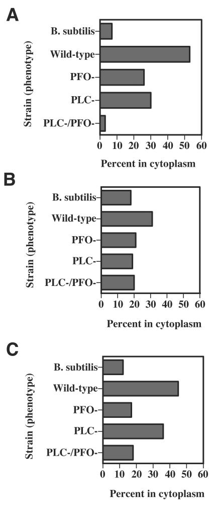
FIG. 6.
(A) Percentage of intracellular C. perfringens bacteria that were determined to be in the cytoplasm of J774-33 macrophages by using electron microscopy (see Materials and Methods). Bacteria clearly lacking a phagosomal membrane around them were scored as being in the cytoplasm of the cells. See Fig. 5A for an example. (B and C) Percentage of intracellular C. perfringens bacteria that were determined to be in the cytoplasm of mouse peritoneal macrophages. Bacteria clearly lacking a phagosomal membrane around them were scored as being in the cytoplasm of the cells. See Fig. 5C for an example. The macrophages in panels A and B were incubated with C. perfringens bacteria for 1 h, while panel C shows results from peritoneal macrophages incubated with the bacteria for 2 h before the cells were fixed and processed for electron microscopy.

