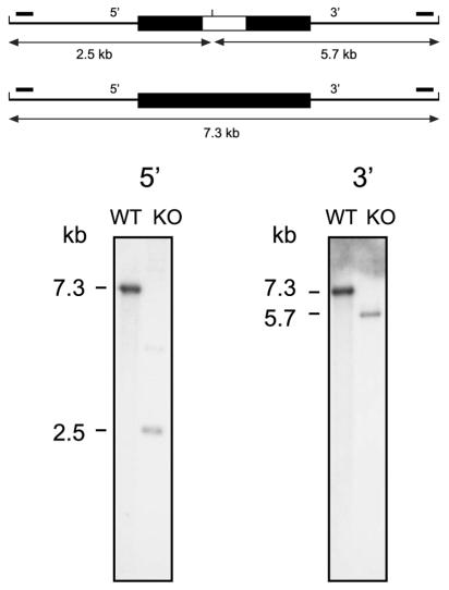
FIG. 1.
Identification of an Rv1931c mutant by Southern hybridization. At the top a schematic representation of the gene locus is shown in which the upper part shows the arrangement in the mutant and the lower part shows that in the wild type. The cloned region is shown as a thick black line, and the introduced kanamycin resistance gene in the mutant is shown as a white box within this. The locations of the hybridizing probes (small solid bars) and the expected sizes of the hybridizing fragments for each of the 5′ and 3′ Southern blots are indicated. Genomic DNAs isolated from the mutant strain (KO) and the parental wild-type (WT) strain were digested with ClaI, transferred to nylon membrane, and hybridized to radioactively labeled DNA probes. The sizes of the hybridizing bands were determined from the migration distances of the DNA molecular size markers λ-HindIII+EcoRI and λ-HindIII.

