FIG. 4.
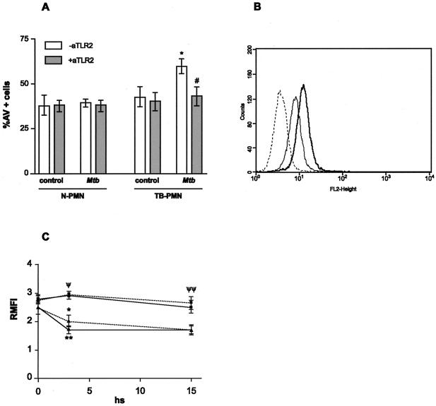
TLR2 triggers apoptosis in TB-PMN. (A) Blocking of TLR2 abolishes M. tuberculosis-induced apoptosis in TB-PMN. PMN (3 × 10⁶/ml) from eight controls and eight TB patients were treated with specific antibody against TLR2 (aTLR2) or its corresponding isotype following incubation with or without M. tuberculosis (Mtb) (106 /ml) for 18 h. Apoptotic cells were evaluated by AV-FITC binding assay as described in Materials and Methods). *, P < 0.001 (M. tuberculosis versus control); #, P < 0.04 (M. tuberculosis versus M. tuberculosis plus anti-TLR2. (B) TLR2 expression in freshly isolated PMN. Expression of TLR2 in TB-PMN () and N-PMN (—) assessed by using an FITC-anti-TLR2 antibody and FACScan. Isotype-control antibody-stained cells are shown as a dotted line histogram. An example from 13 experiments done in each group are shown. (C) TLR expression in N-PMN (▴) and TB-PMN (▪) cultured with () or without (—) M. tuberculosis at 0, 3, and 18 h. Expression of TLR2 was evaluated and expressed as relative MFI as detailed in Materials and Methods. *, P < 0.04; **, P < 0.002 (control versus M. tuberculosis N-PMN) (n = 8). ψ, P < 0.02; ψψ, P < 0.0004 (TB-PMN vs. N-PMN) (n = 8).

