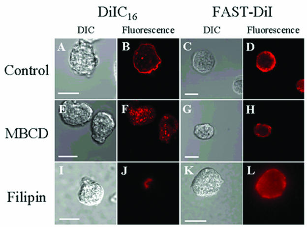
FIG. 1.
Fluorescence microscopy of Entamoeba stained with the fluorescent lipid analogues DiIC16 (raft) and FAST-DiI (nonraft). Both stains were detected in the plasma membrane in untreated control cells and in intracellular structures (B and D). Treatment of cells with the cholesterol-depleting agent MBCD or the cholesterol-sequestering agent filipin resulted in an altered staining pattern for DiIC16 (F and J) but not for FAST-DiI (H and L). Panels A, C, E, G, I, and K represent differential interference contrast (DIC) images. Scale bars, 20 μm.

