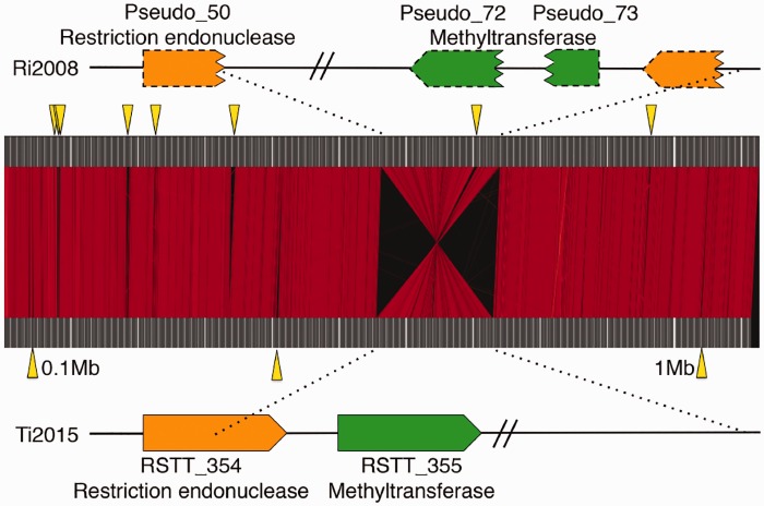
Fig. 1.—
Synteny of chromosomes between genomovars Ri2008 and Ti2015. Upper and lower columns indicate the chromosomes of genomovars Ri2008 and Ti2015, respectively. Red lines show the regions with ≥90% nucleotide sequence identity between the genomovars. Yellow wedges indicate the positions of deletions ≥1kb length. A schematic view of the pseudogenization of an R–M system caused by a large inversion is also shown. A restriction endonuclease gene in Ti2015 (RSTT_354: orange box) was split into two parts by the inversion and found as pseudogene fragments in Ri2008 (two dashed orange boxes). An adjacent methyltransferase gene (RSTT_355: green box) was also split into two and found as pseudogene fragments in Ri2008 (two dashed green boxes). The length of the boxes indicates the relative sequence length. Wavy ends of CDSs indicate split sites. Other genes around the inverted region are not shown, and omitted regions are indicated by double slashes.

