
Figure 1.
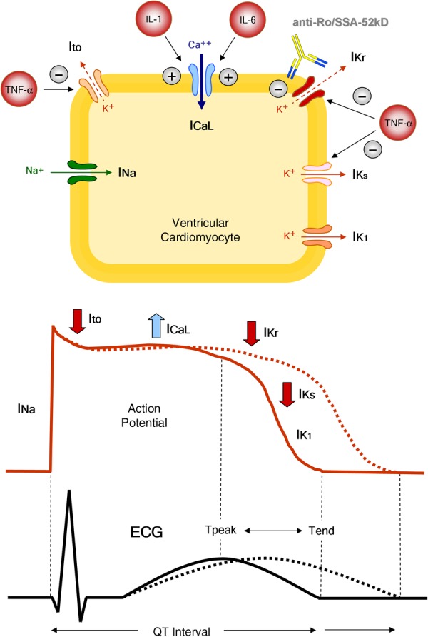
QT interval, inflammation and autoimmunity. From the cell to the surface ECG. (i) Inflammatory cytokines prolong APD and QTc by directly modulating the expression and function of potassium or calcium channels in cardiomyocyte, resulting in changes of the related currents: TNF-α inhibits IKr, IKs and IK1 currents, while IL-1 and IL-6 enhance the L-type Ca2+ current. (ii) Anti-Ro/SSA-52kD directly cross-react with the extracellular loop of the hERG-potassium channel pore-forming region, leading to IKr inhibition with APD and QTc prolongation. APD, action potential duration; QTc, heart rate-corrected QT interval; TNF-α, tumour necrosis factor α; IL-1, interleukin-1; IL-6, interleukin-6; Na+, sodium; Ca2+, calcium; K+, potassium; INa, sodium current; Ito, transient outward current; ICaL, L(long-lasting)-type calcium current; IKr, rapid component of the delayed rectifier potassium current; IKs, slow component of the delayed rectifier potassium current; IK1, inward rectifier potassium current; anti-Ro/SSA-52kD, anti-Ro/SSA-52kD antibodies. Modiified from: Lazzerini et al.4