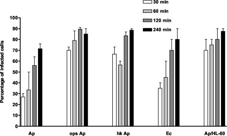
FIG. 2.
Percentages of neutrophils containing intracellular A. phagocytophilum after incubation for 30, 60, 120, and 240 min, determined by using double-labeling immunofluorescence microscopy. Unopsonized A. phagocytophilum (Ap), opsonized A. phagocytophilum (ops Ap), heat-killed A. phagocytophilum (hk Ap), and unopsonized E. coli (Ec) (as a control) are compared. HL-60 cells were also incubated with unopsonized A. phagocytophilum (Ap/HL-60).

