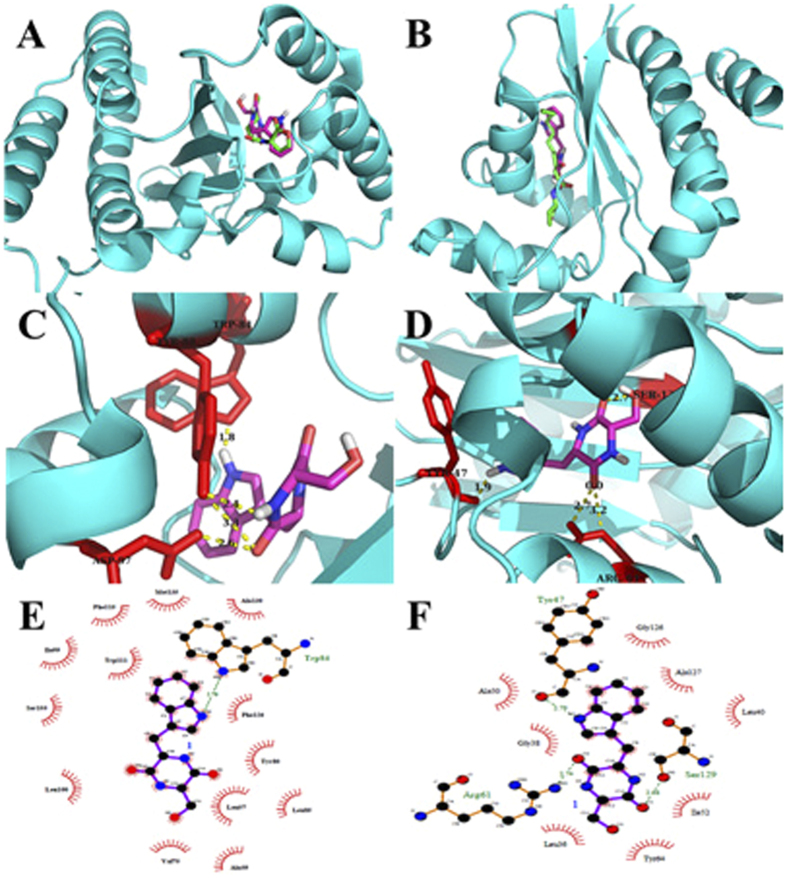
Figure 4.
Docked conformation of natural ligand C6HSL (left) and 3-oxo-C12HSL (right) with green sticks and cyclo(Trp-Ser) with purple sticks into the active site of 3QP1 and 2UV0 receptor protein respectively (A,B); Analysis of docked cyclo(Trp-Ser) bound to 3QP1 (left) and 2UV0 (right) showing the key interactions in the binding pocket. The hydrogen bonds are shown with dotted yellow (C,D); Ligplot of cyclo(Trp-Ser) bound to 3QP1 (left) and 2UV0 (right) showing the key hydrophobic interactions (E,F).

