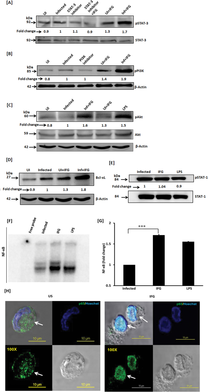
Figure 6. IFG signaling of the macrophages induces phosphorylation of STAT-3 through Akt-PI3K pathway and bolsters NF-κB nuclear translocation.
Mtb infected macrophages were cultured with IFG. The cell lysate (cytosolic extract) was prepared and Western blotting was performed to monitor the expression of [A] pSTAT-3; [B] pPI3K; [C] pAkt; [D] Bcl-xL; [E] pSTAT-1. [F,G] NF-κB was detected using nuclear extract by EMSA and densitometry values are represented by bar diagram as a fold change (mean ± SEM). [H] The translocation of NF-κB from cytosol [US] to nucleus [IFG] was further validated by confocal microscopy using IFG stimulated Mtb infected macrophages. The green fluorescence indicates NF-κB marker p65 and blue signifies the nucleus staining with Hoechst dye (magnification: 100x). LPS is used as a positive control. US: infected macrophages [no IFG]; IFG: infected macrophages treated with IFG. Data are representative of two independent experiments. ***p ≤ 0.0002.

