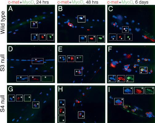
Figure 2.
Expression of c-met and MyoD in fiber-associated satellite cells. After 24 h (A,D,G), 48 h (B,E,H), or 144 h (C,F,I) in culture after harvest, myofibers were fixed and stained for the satellite cell marker c-met (red) and the myogenic regulatory transcription factor MyoD (green); nuclei were visualized with DAPI. (C,D,G) Note that some DAPI-stained nuclei in insets are myonuclei of the parent fiber and not satellite cells, and thus are not positive for any other stains. Wild-type satellite cells (A-C) are activated by fiber harvest and can be distinguished from the host myofiber at 24 h by their morphology and expression of MyoD in some cells (A). They divide synchronously between 24 and 48 h while the percentage of cells expressing MyoD increases (B) and continue to proliferate on the host myofiber (C) as well as in adherent colonies on the culture plate (see Fig. 5). (D-F) Syndecan-3-/- cells displayed grossly normal phenotypes except that MyoD expression is somewhat reduced and is often mislocalized: Inset in E shows two sister satellite cells, one that does not express MyoD and one with appropriate nuclear MyoD staining, whereas all of the satellite cells in F display inappropriate cytoplasmic MyoD staining. (G-I) Syndecan-4-/- cells displayed defective patterns of activation (G), proliferation (H), differentiation, and association (I), as well as a low incidence of MyoD expression (cells in G,H do not express MyoD), coupled with a high level of MyoD mislocalization (none of the clustered cells in I display nuclear MyoD staining; see Fig. 4). Asterisks label satellite cells.

