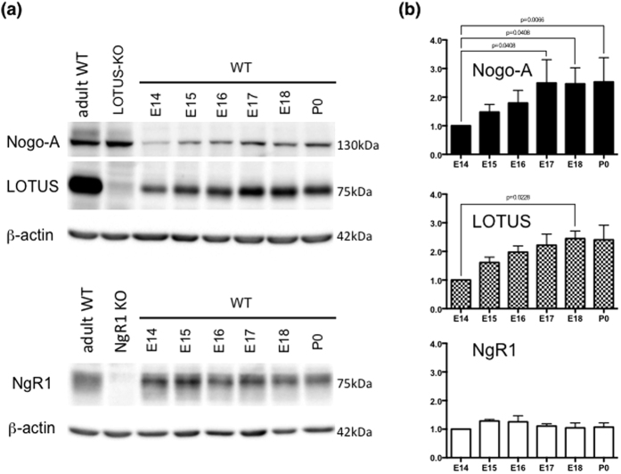
Figure 2. Expression of Nogo-A, NgR1 and LOTUS in the OB.
(a) Immunoblots of Nogo-A, NgR1 and LOTUS in the olfactory bulb at E14, 15, 16, 17, 18 and P0. WT, LOTUS-KO and NgR1-KO indicate protein lysates from wild-type mice at P305, lotus-deficient mice at P237 and ngr1-deficient mice at P91, respectively. β-actin is used as an internal control protein. (b) The expression levels of Nogo-A, NgR1 and LOTUS are quantified by the intensity of each protein immunoblot and normalized to the intensity of β-actin. The significance level was analyzed by performing a Kruskall-Wallis test with Dunn’s multiple comparison analysis. (Nogo-A: n = 4 experiments; LOTUS: n = 3 experiments; NgR1: n = 3 experiments).

