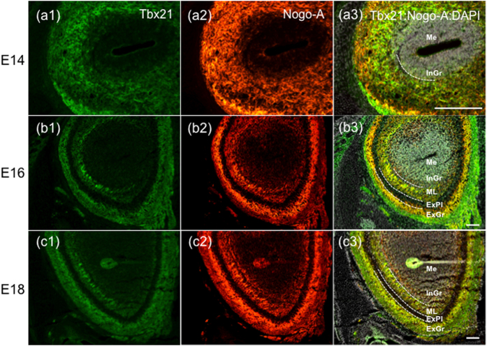
Figure 3. Expression of Nogo-A in mitral/tufted cell layer of the developing OB.
(a–c) Fluorescent immunohistochemistry of Tbx21 (a1,b1,c1), Nogo-A (a2,b2,c2) and their merged image with DAPI (a3,b3,c3) in the developing OB of wild type mice at E14, 16, and 18. Nogo-A and Tbx21 were co-expressed outside the internal granular layer (a3). At E16 and E18, Nogo-A and Tbx21 were co-expressed at the mitral cell layer, the external plexiform layer and the glomerular layer (b3,c3). Scale Bars: 100 μm. Dotted lines are the borders between each cell layer. Me: medulla, InGr: internal granular layer, MiCe: mitral cell layer, ExPl: external plexiform layer, GlLa: glomerular layer.

