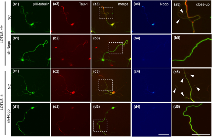
Figure 4. Nogo signaling mediates axonal collateral formation in cultured OB neurons.
(a1–d1) Immunostaining of βIII-tubulin, a neuronal marker. (a2–d2) Immunostaining of Tau-1, an axonal marker in the same cells of (1). (a3–d3) Merged images of (1) and (2) and (a4–d4) immunostaining of Nogo-A in pseudo-color. (a5–d5) Dashed boxes in images of a3–d3 correspond to images of a5–d5 at higher magnification. Arrowheads indicate axonal collateral branches. Dissociated OB neurons from E14.5 wild-type mice (LOTUS+/+) (a,b) or lotus-deficient mice (LOTUS−/−) (c,d) were cultured for 7 days. The OB neurons were infected with lentiviral particles encoding a shRNA sequence (sh1 in b, sh3 in d) against Nogo-A mRNA (sh-Nogo) (b,d) or lentiviral particles encoding a negative control shRNA sequence (NC) (a,c). Compared with that in the LOTUS+/+ mice (NC) (a), axonal branching points were increased (arrowheads in c5) in LOTUS−/− mice (NC) (c), whereas knockdown of Nogo-A (sh-Nogo) resulted in reduction in branching points (b,d). Scale bars: 50 μm.

