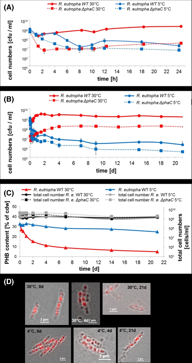
Figure 3.
Viable cell counts, total cell numbers and PHB contents of R. eutropha H16 and R. eutropha ΔphaC cells during incubation in PBS. Cells were grown in NB medium supplemented with 0.2% of sodium gluconate for 5.5 h, harvested by centrifugation and then suspended in PBS and incubated at 30°C (red line) or at 5°C (blue line). Wild type cells (solid lines) had ∼32% accumulated PHB and ΔphaC cells (dotted lines) were free of any storage PHB. At indicated points of time, viable cell counts [cfu ml−1] (A and B), and total number of cells [cells ml−1] and PHB content [% of cellular dry weight (cdw), mean of two determinations] (C) were determined. In (A), the time scale of the first 24 h is enlarged relative to the time scale of 3 weeks in (B). The same log scale of 5 decades is given in all graphs for better comparability. Examples of microscopical images of Nile red-stained wild type cells (overlay of bright field image and fluorescence image) are shown in (D). Note, the decrease in the number of red-stained PHB granules after 21 days at 30°C but not at 5°C. Error bars indicate standard deviation. PHB contents of the phaC mutant were not determined because the inability to synthesise PHB in the absence of PHB synthase has been frequently reported.

