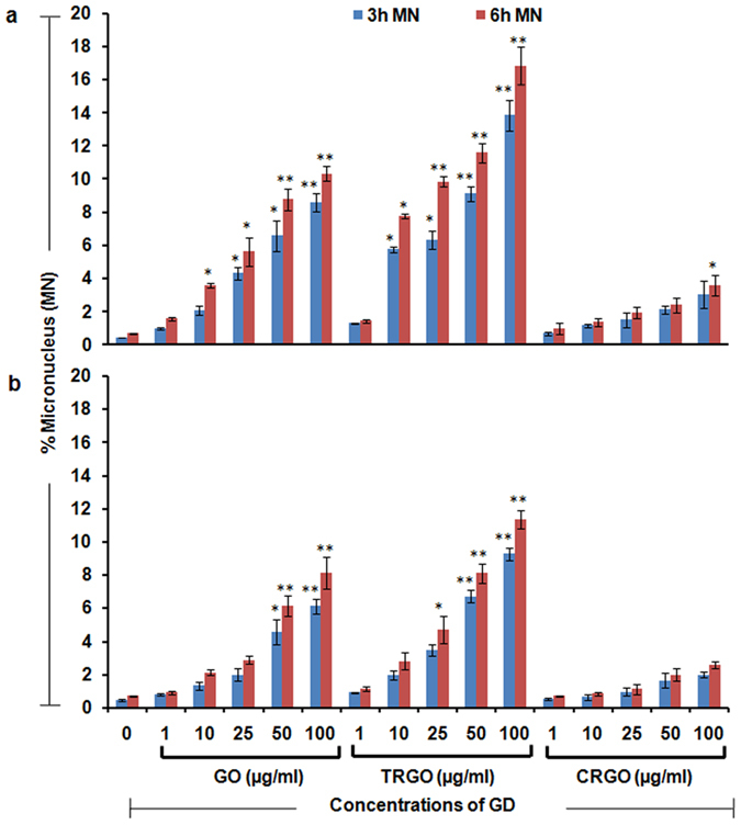
Figure 4. Genotoxicity potential of GD in human lung cells.

Flow cytometry based micronucleus (MN) assay indicate a significant increase in MN upon the exposure of GO, TRGO and CRGO in (a) A549 cells and (b) BEAS-2B cells after 3 h and 6 h time period at a concentration range of 1–100 μg/ml. A notable effect was found in TRGO exposed and more especially A549 cells which showed the DNA damaging potential of GD. Data represents mean ± SE of three independent experiment. *p < 0.05, **p < 0.01, compared to control value.
