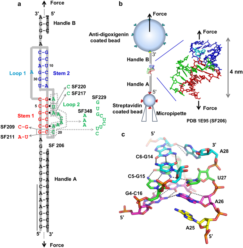
Figure 1. Pseudoknot structures and optical tweezers experimental setup.
(a) Secondary structure of pseudoknot SF206 and mutants. The slippery sequence (G GGA AAC) is indicated by a vertical black line. The DNA strands (highlighted in dark gray) are complementary to the mRNA (highlighted in light gray) forming the handles A and B for the pulling experiments. (b) Experimental setup of single-molecule force spectroscopy using Minitweezers. The NMR structure18 is shown with the same color coding as panel a. The drawing is not to scale. (c) Structures of the three consecutive minor-groove base triples A28∙C6-G14 (cyan), U27∙C5-G15 (green), and A26∙G4-C16 (magenta) found in SF206.

