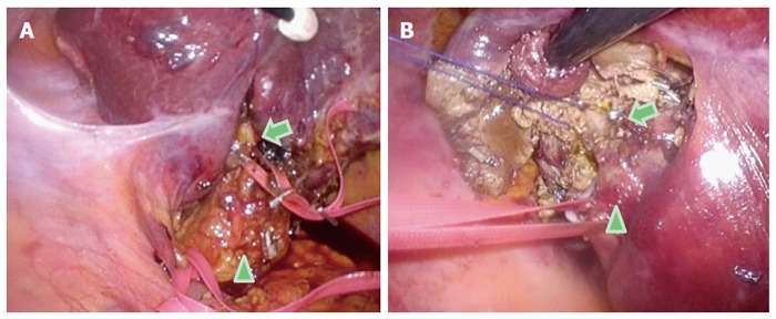
Figure 4.

Intraoperative findings of Glissonian pedicles during the semi-prone position caudal approach for segment 7 segmentectomy. For the segment (S) 7 segmentectomy, a good view and access to the right part of the hilar plate, posterior and the S7 Glissonian pedicles are established upon flipping-up of the liver S6 and of the gallbladder in the left upward direction. A: The arrowhead indicates the hepatoduodenal ligament encircled with tape; the arrow indicates the posterior branch of the Glissonian pedicle encircled with tape in the Rouviere’s sulcus; B: The arrowhead indicates the posterior branch of the Glissonian pedicle encircled with tape; the arrow shows the S7 branch of the Glissonian pedicle encircled with string.
