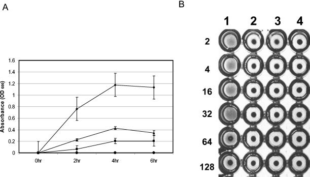
FIG. 3.
Hemolytic and hemagglutinin activities of P. gingivalis FLL93 is reduced. Bacterial cells from overnight cultures were harvested by centrifugation, washed three times with 1× PBS, and then resuspended to a final OD600 of 1.5. Sheep erythrocytes were harvested by centrifugation and washed with 1× PBS until the supernatant was visually free of hemoglobin pigment. The washed erythrocytes were suspended to 1% in 1× PBS. (A) Hemolytic activity was determined by mixing equal volumes of bacterial cells with 1% erythrocytes in PBS. This mixture was then incubated at 37°C. Samples (500 μl) were withdrawn every 2 h and then were centrifuged. The OD405 was determined by spectrophotometry. As a negative control, erythrocytes were used alone. This experiment was done in triplicate, and average values are plotted (diamonds, W83; squares, FLL92; triangles, FLL93; circles, negative control). (B) Hemagglutination activity of P. gingivalis was performed on P. gingivalis W83, FLL92, and FLL93 cells that were serially diluted twofold in 1× PBS. Aliquots (100 μl) of the dilution were then mixed with an equal volume of 1% sheep erythrocytes and were incubated at 4°C for 3 h in a round-bottom microtiter plate. The hemagglutination titer was defined as the last dilution that showed full agglutination. Lane 1, W83; lane 2, FLL92; lane 3, FLL93; lane 4, negative control (sheep erythrocytes alone; Fig. 3B). Error bars indicate the standard error of the mean of three independent trials.

