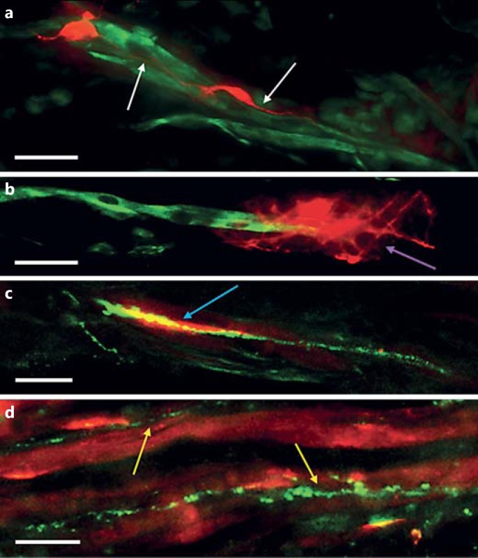
Fig. 2.
Neurite development and synaptic contact within 3D collagen-based co-culture constructs. Longitudinal slices (30 µm) were taken from 3D constructs for immunostaining and imaging. a, b Sections were stained for desmin (green) and MAP-2 (red). Scale bars = 20 μm. c, d Sections were stained for SV-2 (green) and AChRs (red). Scale bars = 10 μm. a Neurites were typically seen tracking along parallel, and in close proximity to, underlying myotubes (white arrows). b Occasionally, neurites were also found wrapped around cultured myotubes (purple arrow). c Pre- and post-synaptic co-localisation (blue arrow) was observed at a frequency of 4.24/mm2. d SV-2 tracks followed underlying myotube orientations (yellow arrows) indicating the path of neurite development and axonal transport of synaptic proteins from the cell bodies toward the developing growth cone.

