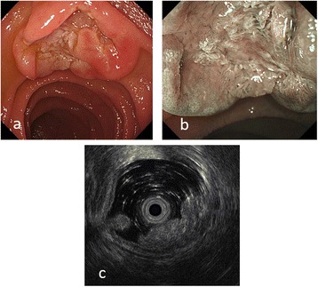
Fig. 1.

a An endoscopic picture showing a wide-based sessile submucosal tumor-like mass with depression at the top of the lesion. b Narrow banding image with magnification showing non-irregular microvessel or mucosal structure. c Endoscopic ultrasound images showing a heterogeneously hyperechoic mass with several anechoic lesions located predominantly in the mucosa
